Localisation of a neurological lesion:
Assuming, where relevant, the patient’s ABCs have been catered to, the crucial step in the management of a patient presenting with symptoms attributable to the nervous system is to localise any neurological lesion. Only then can the possible causes be narrowed down into a manageable few to guide appropriate workup. Have a neuroanatomical schema to guide you, with the location of the main neural functions tested in the neurological examination, including: behaviour and arousal; ascending sensory systems (tactile, proprioception and nociception); motor function (posture and gait); cranial nerve function; and spinal reflexes.
Approach the situation asking five broad questions:
Does this complaint of weakness represent a true loss of motor power?
Because weakness might be described in any number of ways or mean different things to different people, e.g. fatigue, malaise, anhedonia, heaviness, “lack of energy,” even numbness, take time to distinguish discrepancies between the patient’s and your own understanding of what the words they use mean. Ultimately, does the patient’s history convey a true sense of loss of motor power? Interrogate by asking such questions as, were you able to raise up your arm, hold a cup of coffee, open the door? How did you get in the car? What can you no longer do?2
What is the Geography of Weakness? — patterns of motor power loss
Some presentations of motor loss are specifically associated with certain anatomical locations of a lesion so as to immediately help to broadly discriminate between locations:
Paraplegia:
- spinal cord
- peripheral nerves
A paraplegia, i.e. weakness of both legs, suggests either a spinal cord lesion or lesions of peripheral nerves.
Ipsilateral hemiplegia:
- opposite cerebral hemisphere
- face, arm, and leg involvement: subcortical lesion
- face and arm involvement +/- cortical features: cortical lesion
The loss of motor power on one-side of the body suggests a lesion in the opposite hemisphere of the brain: a subcortical lesion (e.g. stroke) if the weakness affects the face, arm, and leg; a cortical stroke if the lesion affects only the face and arm and spares the legs, especially if associated with any cortical symptoms. Hemiplegia can result from a unilateral lesion of the brain stem, internal capsule, or cerebral cortex. Brain-stem lesions result in crossed hemiplegia. For example, a left pontine lesion will result in left facial weakness of lower motor neuron type and right-sided hemiplegia. Similarly, a lesion in the left midbrain will result in left-sided oculomotor weakness with right hemiparesis and right facial weakness of upper motor neuron type. This constellation of signs is called Weber syndrome. Lesions above the level of the brainstem result in uncrossed hemiplegia. For example, a lesion in the left internal capsule would result in right hemiplegia and right facial weakness of the upper motor neuron type. A left cortical lesion may also result in cortical dysfunction in addition to right hemiparesis and facial weakness of upper motor neuron type.2
All 4 limbs (quadriplegia) :
- high spinal (cervical) cord
- peripheral nerve – though unlikely to involve arms and legs simultaneously
- myopathy
Where all four limbs are involved, think of a high spinal (cervical) cord lesion; very occasionally disease of the peripheral nerves or even a myopathy may present that way.
What is the time-course of acute motor weakness, and does it also reflect any fatiguability to that weakness?
Getting a feel for the time-course of events is critical to any clinical enquiry, not least of all the neurological case. How quickly did the patient go from no symptoms to the point where their symptoms had become maximal (i.e. rapidity of onset)?
- Abrupt onset: of power loss is a vascular cause until proven otherwise. Lacunar/small vessel infarcts, however, may present in a stuttering fashion, over minutes or hours.
- Onset over minutes to hours: consider metabolic disturbances or toxins (or a lacunar/small vessel stroke).
- Onset over many hours: consider lacunar/small vessel stroke, Guillain-Barré syndrome, myasthenia gravis and tick paralysis.
- Onset over many days: peripheral neuropathies and neuromuscular junction disease usually take a week or longer to develop
The course of decreased motor strength can also hint at the etiology. Power loss that frequently relapses or fluctuates can be related to multiple sclerosis, periodic paralysis, and myasthenia gravis. On the other hand, transient motor loss may be caused by peripheral nerve entrapment or hemiplegic migraine. Fatigability is also an important consideration in the neurological review. Diminished strength with repeated normal use or testing of the muscle is highly suspicious for a neuromuscular junction disease such as myasthenia gravis.
What associated features are present?
Once a lesion has been localised to a neuroanatomical region, scrutinise the clinical presentation for associated symptoms that will help further discriminate between possible lesion locations:
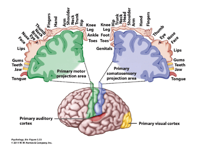


Does the motor power loss represent an upper or a lower motor neuron weakness?
Apart from the increased tone and reflexes (incl. Babinski), an upper motor neuron loss of power also comes with slowness. When a patient is asked to tap their foot or roll their forearms, the tempo is slow in those with a corticospinal tract lesion. In patients with lower motor neuron lesions, their tapping rolling speed is not affected.
Peripheral nerve: this may be suggested by mixed motor and sensory symptoms. In a patient presenting with loss of motor power and numbness, this at least suggests a problem proximal to the neuromuscular junction, either in the peripheral nerve, spinal cord, or above. Sensory pathways consist of the lateral spinothalamic tract and the dorsal column medial lemniscal pathway. Pain and temperature sensations are carried through the lateral spinothalamic tract, while the dorsal column tract carries fine touch, vibration, and proprioception. First-order neurons are located in the dorsal root ganglion of spinal nerves. Neuropathy is a process of axonal degeneration or demyelination of nerves. In neuropathic diseases, sensory involvement usually occurs before motor impairment with the longest nerves being affected first. An example is Guillain-Barre syndrome (GBS) where an ascending paresthesia occurs first, followed by motor weakness.2
NMJ: Neuromuscular junction disorders are usually systemic due to antibodies against acetylcholine receptor. Distinguishing features of neuromuscular junction processes include fluctuating weakness along with early involvement of the ocular (diplopia, ptosis) and bulbar (tongue, jaw, face, larynx) musculature.2
Muscle: Myopathies and neuromuscular junctional pathologies usually involve purely motor deficits. Myopathies are painful, usually symmetric, with proximal muscles involved.2
Summary: five-part schema to assess for weakness
- Does this complaint of weakness represent a true loss of motor power?
- What is the geography of this weakness – patterns of motor power loss?
- Timing, course, and fatigability of acute motor weakness
- Associated findings that help with localisation
- language dysfunction (aphasia): dominant (usually left) hemisphere; or neglect of one side of their body or of the world: non-dominant (usually right) hemisphere
- pure motor syndrome without sensory signs: subcortical lesion
- diplopia, dysarthria, dysphagia: i.e. bulbar (brainstem)
- bladder dysfunction: spinal cord, i.e. if the bladder is spared it is unlikely to be a spinal cord lesion
- absence of reflexes: peripheral neuropathy2
- Distinguish an upper (UMN) from a lower motor neuron (LMN) weakness
Note: Hemi-spatial neglect can be examined through observation and visual field testing. Hemi-sensory neglect can be examined with the double extinction test. Begin with tapping on the patient’s forearm on one side and asking which side they felt, right or left. Then repeat the test on the other side. Then tap on both forearms simultaneously. The patient with hemi-sensory neglect will report sensation only on the non-neglected side.2
A spinal cord syndrome may develop from extramedullary and intramedullary pathological processes. Initial symptoms may be gradual in onset and progressive, including pain, dysesthesia, or subtle upper-or lower-extremity weakness. In other cases, such as an inflammatory myelitis, acute onset of severe motor, sensory, and autonomic deficits may develop without premonitory symptoms. Trauma from a cervical flexion-extension injury, for example, may produce a central cord injury of the lower cervical spinal cord with incomplete quadraparesis, whereas a complete transection injury at the lower thoracic spinal cord from a fall may result in complete paraplegia. Thus, both the rostro-caudal segmental level of disease involvement or trauma and completeness of the lesion in the transverse plane anticipate the person’s impairments and disability. The sensorimotor clinical examination allows localization of the lesion
References
- Assir MZK, M Das J. “How to Localize Neurologic Lesions by Physical Examination.” [Updated 2020 Dec 15]. In: StatPearls [Internet]. Treasure Island (FL): StatPearls Publishing; 2021 Jan-. Available from: https://www.ncbi.nlm.nih.gov/books/NBK493159/.
- Helman, A. Porfiris, G. Baskind, R. “Episode 156-Acute Weakness.” Emergency Medicine Cases. May, 2021. https://emergencymedicinecases.com/acuteweakness. Accessed Aug 8, 2021.
- Helman, A. Baskin, R. Porfiris, G. “Neuromuscular Disease for Emergency Medicine.” Emergency Medicine Cases. June, 2021. https://emergencymedicinecases.com/neuromuscular-disease. Accessed Aug 8, 2021.
- Dobkin, Bruce H. and Havton Leif A. “Paraplegia and Spinal Cord Syndromes.” https://neupsykey.com/paraplegia-and-spinal-cord-syndromes/.
