Preamble
Through a cell-mediated hypersensitivity, the Mycobacterium tuberculous bacillus provokes in the host a chronic granulomatous infection of predominantly the lungs. It is estimated that half the entire world’s population is so infected with the tubercle bacillus, yet most cases of tuberculosis remain asymptomatic. Nonetheless, three million people each year is a toll too high to humanity. But an increased funding attracted after the WHO-declared “global health emergency” in 1993 led to a stabilisation and then reduction in the global disease burden from TB a decade later.
Background
Tuberculosis transmission occurs from person to person through respiratory droplets in proportion to the number of tubercle bacilli in the sputum (inoculum), which in turn relates to the extent of pulmonary disease, and the frequency of cough, so that extensive disease can be highly contagious. Otherwise, household contact for many months is the rule of thumb for transmission of infection. And most patients are non-infectious within 2 weeks of treatment. Extrapulmonary TB carries a negligible risk of transmission.
Surface lipids and water-soluble cell-wall peptidoglycans help the bacterium survive the intracellular milieu of lung macrophages, which activate a non-specific acute inflammatory response leading to regional lymphadenopathy. If the infection cannot be adequately contained by the regional nodes, infection then becomes disseminated through the bloodstream. Nonetheless, at this time, most lesions heal.
Two to eight weeks following this initial inflammatory response, the evasive bacilli can begin to multiply in macrophages before triggering a cell-mediated hypersensitivity response. Lymphocytes, then monocytes, macrophages, and histiocytes are, in turn, recruited to the site of inflammation leading to the formation of the so-called “caseating granulomas” — granulomas that are characterised by a central necrosis. This is a T-lymphocyte dependent antigen-specific response which confers an acquired immunity to the host, and can usually be detected clinically as tuberculin hypersensitivity (Manteaux test).

A chronic tuberculosis infection develops (usually within 3 to 5 years of the primary infection) in only a minority, who do not successfully contain their primary infection. This is most commonly in adolescents and young adults (younger females, older males). Before puberty, on the other hand (infants are the exception to this), the primary lesions almost always heal. The pathophysiology in infants, however, is one of widespread disseminated (miliary) tuberculosis, including meningeal involvement.
Risk factors for infection then are age, famine/poverty and overcrowding, and immunosuppression. Transmission of the bacterium, which is susceptible to UV light, is rare during the day and in areas of adequate ventilation, the most important factor in transmission reduction.
Clinically, we distinguish between primary tuberculosis infection (asymptomatic or non-specific mid/lower zone pneumonitis with lymph node enlargement) and reactivation (secondary) tuberculosis. There are 3 main routes of primary infection: direct spread to the lungs; infection of the tonsillar lymphatic tissue and from there to the neck nodes, and lower ileal infection (including involvement of ileocaecal lymph nodes). Reactivation tuberculosis is the chronic wasting disease characterised by constitutional symptoms of weight loss, low-grade fever, and drenching night sweats.
Reactivation tuberculosis is predominantly a pulmonary infection with a predilection for the superior segments of both the upper and lower lobes, apical disease being the most common. These patients present with a chronic cough and blood streaks in the sputum. Reactivation disease can occasionally be extra-pulmonary — pleural effusion, pericarditis, cervical adenitis, skeletal (“Pott’s disease” of the spine), joints, genitourinary, meningeal, gastrointestinal, and essentially any organ system involvement is possible.
In due course, the tuberculous bacillus has grown increasingly resistant to routine conventional pharmacotherapy. Multi-drug resistant tuberculosis (MDR-TB) is defined as resistance to at least the two key drugs of modern anti-TB regimens — isoniazid and rifampicin.
Natural History of TB
- Infants: miliary TB
- Children: latent TB (primary lesions almost always heal)
- Adolescents/young adults
- latent TB
- chronic TB (usually manifesting within 3 years of the primary infection)
Diagnosis of TB
Think of TB, especially in migrants from high-burden countries (HBCs) and other at-risk populations (e.g. Indigenous Australians). Co-epidemics of TB in immunocompromised people, particularly those with HIV, should alert the clinician to look for these coinfections in the appropriate clinical context.

Testing
Any person with presenting features consistent with TB, with a past history of TB, or with a positive TB screening test should have a CXR. Those with clinical features and a positive CXR should have smear (x 3) and culture.
- Screening Tests
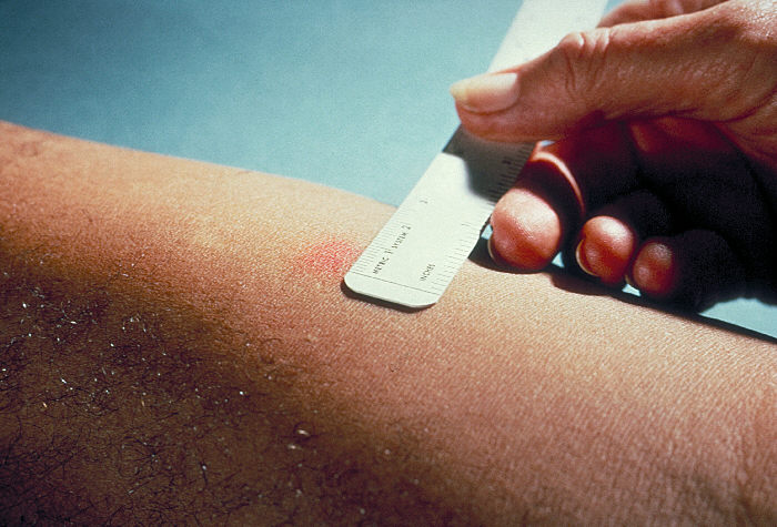
- Tuberculin Skin Test (TST) — Manteaux test
- Interferon-gamma release assay — such as the Quantiferon Gold (QFT-G) test
- Zeil Neilson Stain, Culture, & PCR
- sputum x 3 (induced if necessary), collected 8-24 hours apart, at least one of which is an early morning specimen
- culture for intracellular acid-fast bacilli (AFB)
- produce no pigment
- grow well at 37°C but take 6 weeks
- + PCR of one specimen if symptom positive
- Start by obtaining the least invasive specimen possible for sampling suspected extra-pulmonary sites of disease
- CXR — PA (+ lateral in children)
- Primary Disease
- Secondary Disease
- Latent Infection
- Miliary TB (widespread involvement)
You should then be able to classify the patient into one of three clinical states from which to inform management:
- Latent TB Infection (LTBI) — no disease: TST/QFT-G positive, CXR negative
- INH 10 mg/kg/day (up to 300 mg) for 6 to 9 months (for instance, this treatment is effective in preventing reactivation of TB in HIV positive individuals and also in the under 5-year old close contacts of active disease)
- TB Infection — not clinically active: TST/QFT-G positive, stable CXR (old, healed)
- Old healed, not previously treated TB
- Old healed, previously treated TB
- Suspected/Confirmed Active TB: TST/QFT-G positive &/or CXR positive &/or Sx/signs &/or smear/culture positive
You can then also classify cases in terms of infectivity …
| 0 = negligible infectivity | Extrapulmonary disease |
| 1 = low infectivity | Smear and culture negative pulmonary disease |
| 2 = medium infectivity | Smear negative, culture positive pulmonary disease |
| 3 = high infectivity | Smear positive, culture positive pulmonary disease or culture positive, cavitating pulmonary disease or culture positive laryngeal disease |
An infectious case of active TB is defined as a case scoring 1 or more, and a non-infectious case as one scoring 0.
Management
- Nutrition
- Hygiene
- Triple therapy orally for at least 3 weeks may have sufficed 20 years ago, but today a five-drug regimen is usually employed, for not less than six months, in susceptible TB:
- Rifampicin 600 mg/d (watch for hepatotoxicity)
- Isoniazid 300 mg/d (watch for peripheral neuritis)
- Streptomycin 15 mg/kg daily or two or three times weekly (dizziness, vertigo)
- Pyrazinamide 150-200 mg/d (avoid in gout)
- Ethambutol (watch for visual impairment)
INH and Rifampicin alone are continued to a total of six months

- Examine sputum monthly until clear
- Extra-pulmonary TB may require a prolonged 9-month course
- bedaquiline (2013) and delamanid (2014)
Note: often impossible to eradicate every tubercle bacillus from the body, often lying dormant enveloped in fibrous tissue from which they can potentially reactivate
In general, a patient with pulmonary TB who complies with therapy and does not have drug resistant disease should become non-infectious after two weeks of appropriate anti-tuberculous therapy.
Fast Facts
- 190 000 people died of MDR-TB (2014 estimate)
- “TB mortality has fallen 47% since 1990, with nearly all of that improvement taking place since 2000.”
- 6 million reported new cases (WHO, 2014) of 9.6 million estimated new cases of which an estimated 480,000 were MDR-TB
- MDR-TB in an estimated 3.3% of new TB cases and 20% of previously treated cases
- 1.7 million deaths (2006)
- Culture positive, smear positive, cavitating pulmonary disease is highly infectious
- Rapid tests for TB are now available: Xpert MTB/RIF; GeneXpert Omni; Xpert Ultra
- New drugs, such as TBA-354, are in advanced phases of clinical development
- 15 vaccine candidates are in clinical trials
References
- MJA 2007;186:240-42
- Tuberculosis. Medical Observer. 10 October 2008
- Tuberculosis: Part 2. Medical Observer. 11 March 2011
- Global Tuberculosis Report, 2015 (20th Edition). World Health Organisation.
- Division of Global Migration and Quarantine, National Centre for Emerging and Zoonotic Infectious Disease, CDC, U.S. Department of Health and Human Services. Guidelines for Screening for Tuberculosis Infection and Disease during the Domestic Medical Examination for Newly Arrived Refugees. April 16, 2012
- Tuberculosis – air travel for patients with TB: Guidelines for GPs. Department of Health, State Government, Victoria
Featured Image
Cavitatory Tuberculosis [Wikimedia Commons]