Physiology

Pathways and Processes in Humans

 Acid-Base

Acid is any molecule that can cleave off (Arrhenius) or donate (Brönsted) H+. Base is au contraire molecule that can cleave off OH or accept H+. Acids and bases undergo metabolic conversion (e.g. lactate to glucose in gluconeogenesis, lactate to pyruvate and oxidation in cardiomyocytes), or excretion from the body. The human organism daily produces great quantities of acids – source of protons. These are volatile (respiratory) e.g. carbonic acid or non-volatile (metabolic) organic (lactic acid, fatty acid, ketone bodies, etc.) and inorganic acids (sulphuric acid, phosphoric acid, etc.).

The body maintains acid-base balance in the face of this acid load by various mechanisms:

  1. a chemical buffering system which deals instantaneously with pH changes
  2. a respiratory system reacts quickly (1-3 minutes) to eliminate carbonic acid through exhalation of carbon dioxide (hence, volatile)
  3. a more complex response over hour to days rendered by the kidneys
  4. detoxification of ammonium in the liver
  5. alteration in myocardial substrate utilisation

figure1

Main chemical buffers
Bicarbonate converted back to CO2 and eliminated by the lungs.
image
Carbonic anhydrase in the kidney converts the dissolved CO2 into carbonic acid
Excretion of acid and ways to jeopardize the system.1. A strong non-volatile acid HA dissociates to release H + and poses an immediate threat to plasma pH.2. Bicarbonate buffers the H + and generates CO 2, which is expelled in the lungs and results in depletion of body HCO 3 -. Non-bicarbonate buffers (collectively referred to as B) carry the H + until the kidneys excrete it.3. The kidneys split CO 2 into H + and HCO 3 – and selectively secrete H + into the lumen and HCO 3 – into the blood. In addition, any excess H + from the body fluid is also excreted.4. Most H + excreted in the urine is carried by urinary buffers (UBs).5. Some organic anions (A) (e.g. lactate, ketoanions) can be metabolized to regenerate the HCO 3 -. If A is not metabolizable (e.g. phosphate or sulfate), it is excreted in the urine.* Two possible ways by which metabolic acidosis can occur. (Pham et al, 2015)
Charge balance in human plasma. SIDa, apparent strong ion difference; SIDe, effective strong ion difference; SIG, strong ion gap [Kaplan and Frangos, 2004]. Conforming to a Brønsted–Lowry definition, adopt the Stewart approach of physical chemistry. This is based on charge (redox) interactions in human plasma, the independent control mechanism for pH determination.
 
The most controversial feature is the designation of pH and bicarbonate concentrations as dependent variables, answerable exclusively to three independent variables. These are the strong ion difference (SID), the total concentration of non-volatile weak acid (ATOT), and PCO2. (Aspects of this assertion [however] conflict with traditional renal physiology, and with current models of membrane H+/base transporters, oxidative phosphorylation, and proton and bicarbonate ionophores.)¹   
 
Reference ranges for blood gas results

This approach leaves three parameters to assess:

  • pCO2
  • weak acids (mostly albumin)
  • strong ion difference (SID)

Cellular Respiration – overview

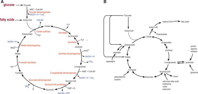
Acetyl-CoA stands at the crossroads of cellular metabolism:

749cd-generalized252520metabolic252520pathway

Glucose metabolism

Glycolysis

Visual representation of glycolysis showing all three phases | Science  nerd, Biology, Chemistry

Oxidation of Pyruvate

Krebs cycle

The citric acid cycle (CAC). A, An overview of CAC reactions. Observe that apart from 3 irreversible steps (citrate synthase, isocitrate dehydrogenase, and 2-oxoglutarate dehydrogenase), the rest of the CAC reactions are reversible, allowing reverse flux under certain conditions (such as in anoxia). B, Anaplerosis and cataplerosis of the CAC. Anaplerosis replenishes CAC intermediates, whereas cataplerosis allows production of various biosynthetic precursors making the CAC as a central metabolic hub, connecting diverse metabolic pathways. CoA-SH indicates reduced coenzyme A; FADH2, reduced flavin adenine dinucleotide; GTP, guanosine triphosphate; NAD+, oxidized nicotinamide adenine dinucleotide; NADH, reduced nicotinamide adenine dinucleotide; and QH2, reduced coenzyme Q. (Czibik et al, 2014)

Oxidative Phosphorylation

ATP synthesis in mitochondria depends on an electrochemical gradient across the mitochondrial inner membrane arising, ultimately, from the energy of reduced electron carriers, NADH and FADH2 (Image: Draw it to Know It)

Pentose Phosphate Pathway

The pentose phosphate pathway provides reduced NADPH and pentose sugars for nucleotide synthesis:

Bryan Krantz: University of California, Berkeley

Protein Metabolism

Structure of amino acids

6b2e6-igen3_06-01_figure-lsmc

Peptide linkage

File:Peptide Bond Formation.jpg

Instantiation of a polypeptide structure

Polypeptide (protein) folding

A three-part diagram shows the generic chemical structure of an amino acid (top), the generic chemical structure of a polypeptide (middle), and the idealized structure of a polypeptide chain folded to form loops (bottom). A dotted line between amino acids on different loops represents their interaction.

Protein structure folding stages - LadyofHats, commons.wikimedia.org

Heme metabolism

Heme-containing proteins

  • Haemoglobin
  • Myoglobin
  • Cytochromes
  • Catalase
  • some peroxidases

Most hemoglobin degradation occurs in the macrophages of the spleen, where globin and iron are conserved for reuse:

image
The globin and iron portions are conserved and reused. Heme is reduced to bilirubin, eventually degraded to urobilinogen, and excreted in the feces. Thus, indirect indicators of erythrocyte or erythrocyte destruction include the blood bilirubin level and urobilinogen concentration in the feces. (Thibodeau GA, Patton KT: Anatomy & physiology, ed 6, St Louis, 2007, Mosby, p 654.)
Heme Degradation
https://microbenotes.com/heme-degradation/

Nucleosides and Uric Acid

Lipid Metabolism

Lipoproteins

Figure 26.16. Schematic Model of Low-Density Lipoprotein.
Schematic Model of Low-Density Lipoprotein (Berg JM, Tymoczko JL, Stryer L., 2002)

Cholesterol metabolism

Cholesterol is primarily stored in the plasma membrane. Upon hormonal stimulation there is increased cholesterol absorption through the plasma membrane. When cholesterol is imported into the cell via the plasma membrane it greatly increases the cholesterol content stored elsewhere in the cell. (Rone, 2009).

Figure 2. Schematic overview of the metabolism of the plasma lipoproteins and reverse cholesterol metabolism. The image was originally published in Atlas of Heart Diseases: Atherosclerosis by Brewer B.H.16 and is reprinted here with kind permission from Current Medicine Group LLC.
Schematic overview of the metabolism of the plasma lipoproteins and reverse cholesterol metabolism

Fatty acid metabolism

Figure 22.2. Steps in Fatty Acid Degradation and Synthesis.
Steps in Fatty Acid Degradation and Synthesis (Berg JM, Tymoczko JL, Stryer L., 2002)

Steroid synthesis

f0f42-21p49
Cholesterol is the sole precursor of steroids. Steroid synthesis is initiated at the inner mitochondrial membrane (IMM), where the cytochrome P450 cholesterol side chain cleavage enzyme (CYP11A1) catalyzes the conversion of cholesterol to pregnenolone. (Rone, 2009)

References


Check my guide for creators: strategies, stories, and tools to help you grow your craft.